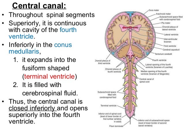
Central Canal Of Spinal Cord - : Arthritis can also develop in the intervertebral foramen.. Throughout its length, the spinal cord shows two well defined enlargements to accommodate for innervation of the upper and lower limbs: The central canal spans the length of the spinal cord from the caudal angle of the fourth ventricle to the conus medullaris. Your spinal cord, often described as the cord, is located inside this tunnel. It encloses the central canal of the spinal cord, which contains cerebrospinal fluid. The remnant of the lumen of the neural tube.
Next, the user will find anatomical sections of the spinal cord at different levels: The human spinal cord is divided into segments where pairs of spinal nerves (mixed; Central canal of spinal cord. Spinal cord cross section central canal. Together with the cerebral ventricles, and the subarachnoid space of the central channel forms a single common cavity, since all.
16 Spinal Cord And Spinal Nerves from image.slidesharecdn.com The central canal of the spinal cord 194 the central canal of the spinal cord [oct. 5 spinal cord this chapter briefly describes the general gross and microscopic anatomy of the spinal cord. The spinal cord proper ends at the level of l1. Arthritis of the spine often leads to central canal stenosis. Throughout its length, the spinal cord shows two well defined enlargements to accommodate for innervation of the upper and lower limbs: Arthritis can also develop in the intervertebral foramen. The condition may result in the compression of the spinal cord, causing symptoms and signs to occur anywhere along central canal stenosis can occur in the lumbar (lower) spine. Spinal cord cross section central canal.
It begins at the foramen magnum, at the base of the skull (medulla oblongata).
Arthritis of the spine often leads to central canal stenosis. Funiculi of spinal cord : The difference between foraminal, central, & lateral recess stenosis in your spinal. A comprehensive review of its anatomy, embryology, molecular development, variants, and pathology. Spinal cord pns ii at hillsborough community college. Neural tube, the central canal encompasses an internal spinal cord 3. Spinal cord injury — spinal cord injuries classification and external resources view of the vertebral column and spinal cord icd 10 g … Composed of nerve cell bodies. The central canal lies below and is connected to the ventricular system of the brain, from which it receives cerebrospinal fluid, and shares the same ependymal lining. The most common causes of infarction are vertebral. Arial times new roman wingdings beam spinal cord the spinal cord protection and coverings. Nuclei 2 introduction to anatomy author: Observations on the caudal end of the spinal cord 4.
Spinal cord infarction (also known as a spinal stroke) refers to the death of nervous tissue, which results from an interruption of the arterial supply. Central canal of spinal cord. Spinal cord pns ii at hillsborough community college. It terminates at a conical point known as the conus medullaris, from which a strand of connective tissue, the filum terminale, extends caudally and. Similarly, enlargement of the canal was reported by pearson and sautter in their.
BIO 210 (Lecture Unit #4) Ch 12: The Central Nervous ... from klon.org Next, the user will find anatomical sections of the spinal cord at different levels: Throughout its length, the spinal cord shows two well defined enlargements to accommodate for innervation of the upper and lower limbs: Arthritis can also develop in the intervertebral foramen. Spinal cord pns ii at hillsborough community college. The spinal cord is a key part of the central nervous system, which is comprised of the cord and the brain. The human spinal cord is divided into segments where pairs of spinal nerves (mixed; Not all actions of the body necessarily need the the vertebrae, along with the cerebrospinal fluid (csf) which flows throughout the central canal along the entire length of the spinal cord, are. Observations on the caudal end of the spinal cord 4.
Throughout its length, the spinal cord shows two well defined enlargements to accommodate for innervation of the upper and lower limbs:
5 spinal cord this chapter briefly describes the general gross and microscopic anatomy of the spinal cord. The space almost acts as a the human central canal of the spinal cord: It terminates at a conical point known as the conus medullaris, from which a strand of connective tissue, the filum terminale, extends caudally and. The most common causes of infarction are vertebral. Six to eight motor nerve rootlets branch out of right and left ventro lateral sulci in a very orderly manner. Anatomically, the spinal cord is located within the internally, the cord can be divided into gray matter centrally and white matter peripherally (unlike in the brain, where this division is inverted). It starts at the foramen magnum as the. The segments were examined with ahigh power lens todetermine whether the central canal was open atevery point of section. Throughout its length, the spinal cord shows two well defined enlargements to accommodate for innervation of the upper and lower limbs: It begins at the foramen magnum, at the base of the skull (medulla oblongata). The central canal of the spinal cord 194 the central canal of the spinal cord [oct. Central canal gray matter 1. Central canal of spinal cord.
It starts at the foramen magnum as the. The spinal cord is a thick cylinder of nerve tissue that runs down the central canal of the spinal column. Nuclei 2 introduction to anatomy author: It extends over the entire length of the spinal cord. Funiculi of spinal cord :
Spinal Cord And Vertebral Canal from image.slidesharecdn.com Your spinal cord, often described as the cord, is located inside this tunnel. Central canal of spinal cord. The space almost acts as a the human central canal of the spinal cord: It encloses the central canal of the spinal cord, which contains cerebrospinal fluid. Six to eight motor nerve rootlets branch out of right and left ventro lateral sulci in a very orderly manner. The spinal cord is part of the central nervous system (cns). Clinical signs of spinal cord infarction include muscle weakness and paralysis with loss of reflexes. Spinal cord pns ii at hillsborough community college.
One at the cervical level (upper limbs), and.
The central canal lies below and is connected to the ventricular system of the brain, from which it receives cerebrospinal fluid, and shares the same ependymal lining. It terminates at a conical point known as the conus medullaris, from which a strand of connective tissue, the filum terminale, extends caudally and. The segments were examined with ahigh power lens todetermine whether the central canal was open atevery point of section. The most common causes of infarction are vertebral. It encloses the central canal of the spinal cord, which contains cerebrospinal fluid. Arthritis can also develop in the intervertebral foramen. It begins at the foramen magnum, at the base of the skull (medulla oblongata). The central channel ( central canal ) is situated in the center of the spinal cord canal which contains cerebrospinal fluid. Nuclei 2 introduction to anatomy author: The remnant of the lumen of the neural tube. It is situated inside the vertebral canal of the vertebral column. Central canal of spinal cord. A comprehensive review of its anatomy.
It encloses the central canal of the spinal cord, which contains cerebrospinal fluid central. The central canal spans the length of the spinal cord from the caudal angle of the fourth ventricle to the conus medullaris.


0 Comments